
With new advances in technology come increasingly innovative ways to improve the quality of life and care. The digital age has changed the way people move through life, the way communities are governed and the way medical professionals diagnose our healthcare issues.
Praveen Rao, Ph.D., is working with a team of pathologists to find a way to automate the everyday tasks of pathologists so that they can focus more on customizing treatments for patients.
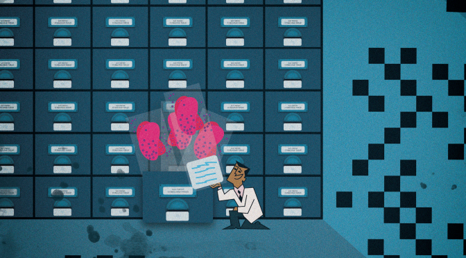
Rao’s work centers on a concept called Whole Slide Imaging, a fairly new wave for digital pathology in the United States. These imaging scanners, which were only approved by the Food and Drug Administration in 2017, will allow pathologists to scan and project tissue slides on screen and use computer algorithms to pinpoint which regions of the slide are important or contain cells of interest. The pathologist can then focus on those specific regions for examination and diagnosis, rather than spending their time manually reviewing each slide.
But what if there was a way for the machine to not only store digital scans — they’re significantly large in size and currently there’s no way to store files digitally — but also to automatically detect cellular and mitotic activity?
That’s what Rao and his team are working on: new techniques to distribute data from whole slide images. They’re building a scalable storage system that will allow different parts of the slide image to be quickly assessed. The National Science Foundation has provided funding for the team to explore commercializing their system. They also submitted a tier one UM System proposal for additional project funding.

As of now, only two companies sell their scanners to hospitals for pathologists to digitize tissue slides and use them for primary diagnoses. Even with digital pathology, Rao says federal law still requires slides to be physically stored for several years, in case doctors need to refer back to them. The storage system Rao and his team are working on will consist of a cluster of machines working together to store and manage data. The goal is to eventually be able to discard physical slides, archive the whole slide image and add new images as they are collected — all using Rao’s storage system.
“Our team is working to apply deep learning algorithms to the images for a good storage system,” Ray says. “In exploring the commercialization of our product, we argue that this platform can be used for next-generation analytics, which we are also working to build so we can package it all together.”
They’re hoping that, eventually, pathologists will be able to look at a patient’s tissue and make a conditional diagnosis, then send the DNA for genomic sequencing. When the DNA comes back it, should help doctors customize treatment based on what a patient’s genetics tell them work best to treat that particular individual.
“If a computer algorithm can predict things accurately, then it relieves pathologists from things like having to manually count how many cells in a particular region have undergone transformations of classifying cells by looking into the microscope,” Rao says. “The more we automate and let the computer do its job, the faster and more scalable it’ll be for a doctor to prescribe customized solutions.”
Quick facts
- Digital pathology is regarded as one of the most promising avenues of diagnostic medicine
- Digital pathology’s market worth is expected to reach nearly $756.1 million by 2022
- Countries currently using digital pathology methods for diagnoses include Canada, the Netherlands and the United Kingdom.
Benefits of digital pathology
- Speeding and simplifying access to histopathology information
- Helping staff work more efficiently
- Centralizing teamwork
